When it comes to an eye exam, most patients think of updating their glasses and contact lenses. While this is true, many overlook what could be the most important thing about their annual exam, eye health! The eye is complex and it provides insight into the health to the entire body, which makes careful examination extremely important.
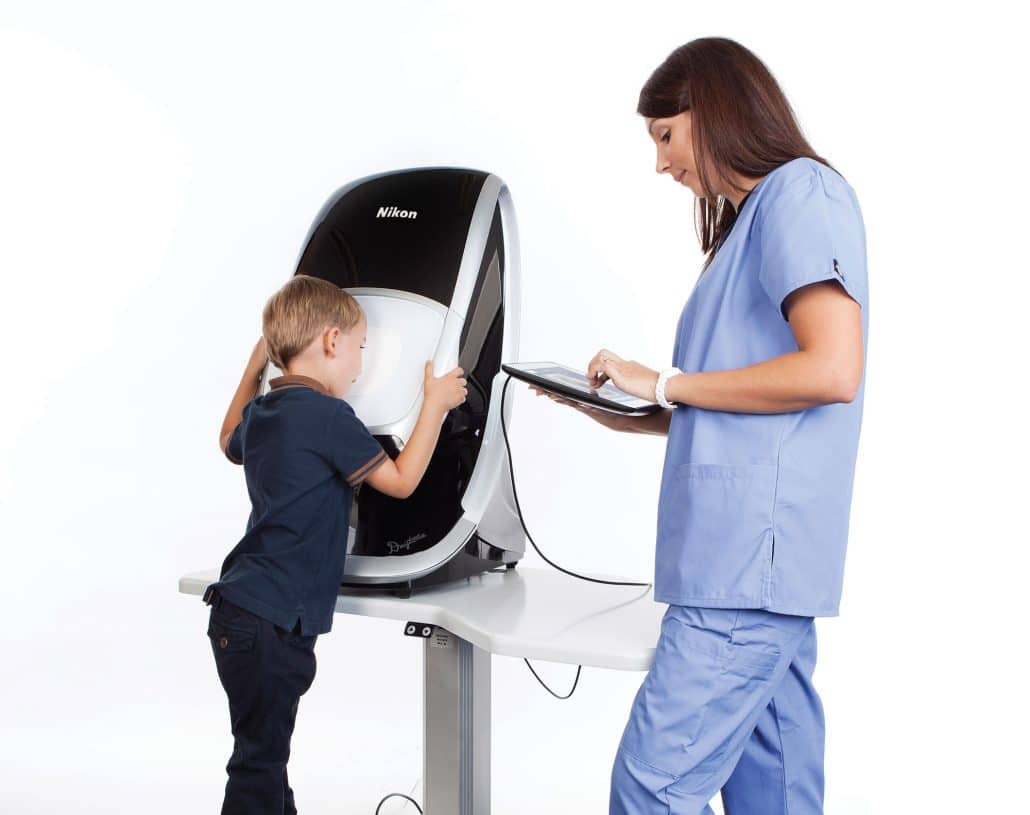
Interesting Eye Fact: The inside of the eye (retina) is the only place in the human body where blood vessels can be seen directly!
By examining the blood vessels inside of the eyes, the doctors can check for many systemic conditions such as diabetes, hypertension, high cholesterol, stoke, anemia, and heart disease – just to name a few. For this reason, we are upgrading every comprehensive eye exam to include optomap digital retinal imaging.
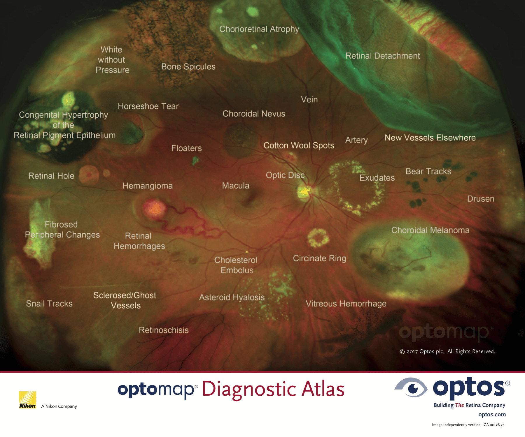
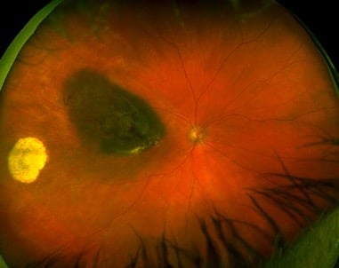
Below is a diagram of some of the things that can be detected on optomap digital retinal imaging:
10 Benefits of Optomap Digital Retinal Imaging:
- Expansive
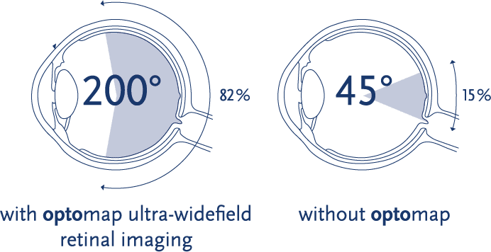
Using a non-invasive spinning camera system, optomap captures an ultra-wide 200 degree view inside of the eye, encompassing 82% of the retina in one image. Additionally, the technicians are trained to modify obtain even father views by taking two or three images if an even wider view is needed.
Traditional imaging methods typically only show 15% of the retina at one time (see image)
- Detail
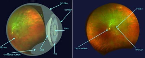
Optomap captures and expands small retinal details onto the full-size computer monitor. For example, the optic nerve is less than 2mm in size, roughly the size of the head of a pin. The imaging technology can magnify the image of the nerve up to the size of a baseball on the screen for the doctor and patient to view in bigger detail. This enhances the ability to gather, monitor, and explain critical information about your eye health.

- Convenience
The comprehensive eye health screening capabilities of optomap will allow most patients to avoid the dilation test. This means most patients can say goodbye to the side effects of blurry vision and light sensitivity that dilation causes, allowing patients to drive home or return to work with better vision immediately after their exam.
*A small percentage of patients will still require dilation at the doctor’s discretion.
- Speed
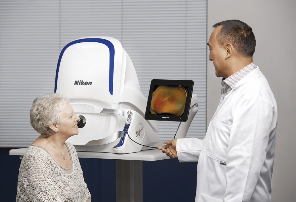
While it takes about 30 minutes for the eyes to dilate, it takes less than 1 second to capture an optomap digital retinal image. Since most patients will not require dilation, this allows for a faster, more predictable visit to the eye doctor.
![]()
- Comfort
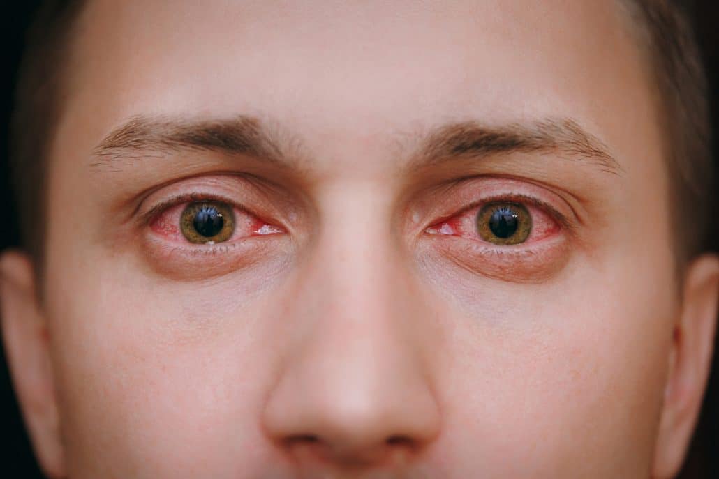
No eye drops are required for optomap retinal imaging. This is welcomed news for patients of all ages who dread the stinging side effect of the dilation drops.
- Patient Education
A photo is worth a thousand words. Complex issues and structural changes become much easier for patients to understand because they can see exactly what the doctor sees while the doctor explains the images to the patient in real time.
- Photodocumenation for the Future
With an annual optomap retinal image, doctors can compare images year after year to better detect subtle changes in the back of the eye. These photographic records can also be shared electronically with patients and other providers when needed.
- Social Distancing
Dilation requires patients to move back and forth between the waiting area and the exam room twice during their eye exam. It also requires a 30 minute wait in the office while the eyes are in the process of dilating. By providing everyone with an optomap retinal image, it reduces the amount of two-way traffic in the office as well as the number of patients sitting in the waiting area. This helps tremendously in creating an environment of enhanced social distancing.
- Sanitary
After each patient, the optomap retinal imaging device is thoroughly cleaned and sanitized by the trained technicians.
- Cutting edge technology
Optomap retinal imaging software contains many special features to make disease detection, patient education, and tracking changes over time fast and easy. Make sure to ask your doctor to review the 3D animation feature and different imaging filters with you on your next appointment!
* several of the images used above are the property of optos*


